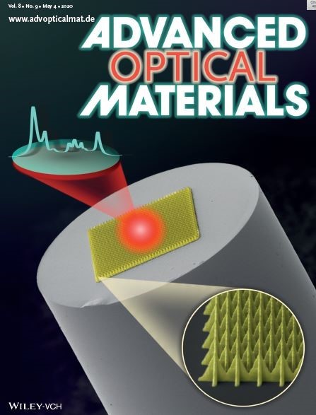


Guang-Zhong Yang’s team Published Advanced Optical Materials Cover Story
May 11, 2020
Professor Guang-Zhong Yang, Dean of the Institute of Medical Robotics, Shanghai Jiaotong University, published the cover story in Advanced Optical Materials as corresponding author, titled “Fiber-Optic SERS Probes Fabricated Using Two-Photon Polymerization for Rapid Detection of Bacteria” on May 4, 2020.

This study presents a novel fiber-optic surface-enhanced Raman spectroscopy (SERS) probe (SERS-on-a-tip) fabricated by simple, precise, and effective two-photon polymerization(2PP). 2PP was adopted to acquire SERS arrays on end-facets of optical fibers. For the SERS-on-a-tip probes, a limit of detection of 10−7 M (Rhodamine 6G) and analytical enhancement factors of up to 1300 are obtained by optimizing the design, geometry, and alignment of the SERS arrays on the tip of the optical fiber. Furthermore, strong repeatability and consistency are achieved for the fabricated SERS arrays, demonstrating that the technique may be suitable for large-scale fabrication procedures in the future. Finally, rapid SERS detection of live Escherichia coli cells is demonstrated. Moreover, it represents the first report of detection of live, unlabeled bacteria using a fiber-optic SERS probe.
When excitation light transmitted through the optical fiber is incident on the SERS array, surface plasmon polaritons (SPPs) are excited and SERS hotspots are generated at nanogaps and nanotips in the SERS array designs. The highly confined electromagnetic fields of these SERS hotspots act to enhance the Raman signal, which is then collected by the optical fiber and delivered to a spectrometer for detection. Raman spectroscopy and infrared spectroscopy belong to molecular vibration spectrum, which can reflect the characteristic structure of molecules. Raman spectroscopy and SERS technology have been widely used in surface science, biomedicine, and etc., They have grown into a powerful analytical tool.
In order to optimize the 2PP process and the SERS performance, the study explored different arrays of nano/microstructures from 2D to 3D structures and optimized the hexagon spacing distance in details. Based on the momentum conservation between the SPP wave vector and the diffraction wave vector, it is possible to theoretically calculate the expected optimal spacing distance and verify it in experiment, so that SERS array can exhibit the best Raman signal enhancement and sensing performance. SEM, the optical microscope images and Raman mapping screening demonstrated the advantages 2PP has in fabricating hexagonally arranged SERS arrays, for example, strong repeatability and consistency. Two common approaches used to create SERS hotspots include chemical synthesis and traditional nanofabrication. The former involves chemical reduction of metal ions, followed by growth and decoration of NPs chemically in solvents. Photolithography, electron-beam lithography, and focused ion beam (FIB) milling are examples of nanofabrication techniques used to produce desired nanostructures and nanopatterns on silicon wafers. Despite being well established, several challenges remain in these methods especially in terms of structure uniformity, efficiency, controllability, repeatability, and complexity of fabrication processes. Moreover, these methods often use toxic chemicals that may leave residual toxins, thus requiring extra purification steps before medical application.
2PP is a mask-less microscale 3D printing technique with sub-diffraction limit resolution down to several nanometers. It is very suitable for micro-scale substrate fabrication, complex micro-structures on end-facets of optical fibers in particular. Accordingly, 2PP has rapidly gained popularity in areas such as micro-optics and photonics, microfluidics, micromechanics, and micro-robotics. This paper’s highlight lies in the first rapid (2.5 ms and 1 s) SERS detection of live, unlabeled Escherichia coli using fused silica planar substrate and fiber-optic SERS probes. It showed a promising application future of new fiber-optic probes in diagnostics.
Full text link:
https://onlinelibrary.wiley.com/doi/10.1002/adom.202070035